These tomographic reconstructions are part of my Palaeobiology MSc project at the University of Bristol, UK. I am extremely lucky of having two great supervisors: Imran A. Rahman (university of Bristol) and Christopher B. Cameron (Université of Montréal). I use a free software (that is right, free!) called SPIERS (http://spiers-software.org). Do not forget to check out the three videos and the gallery below.
Material and Methods
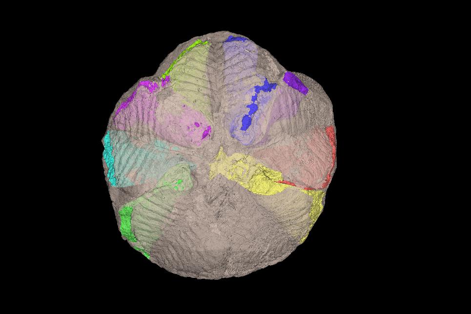
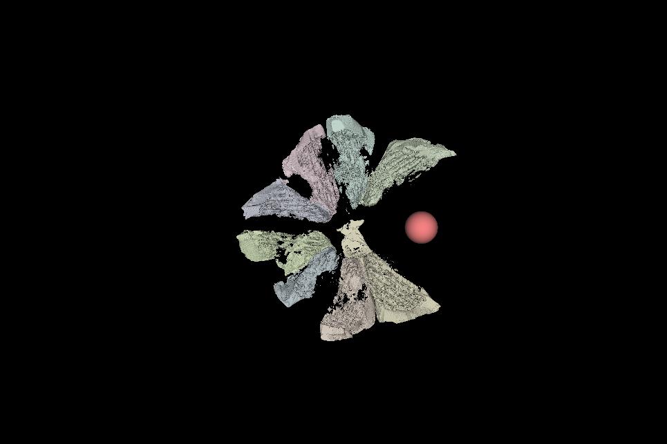
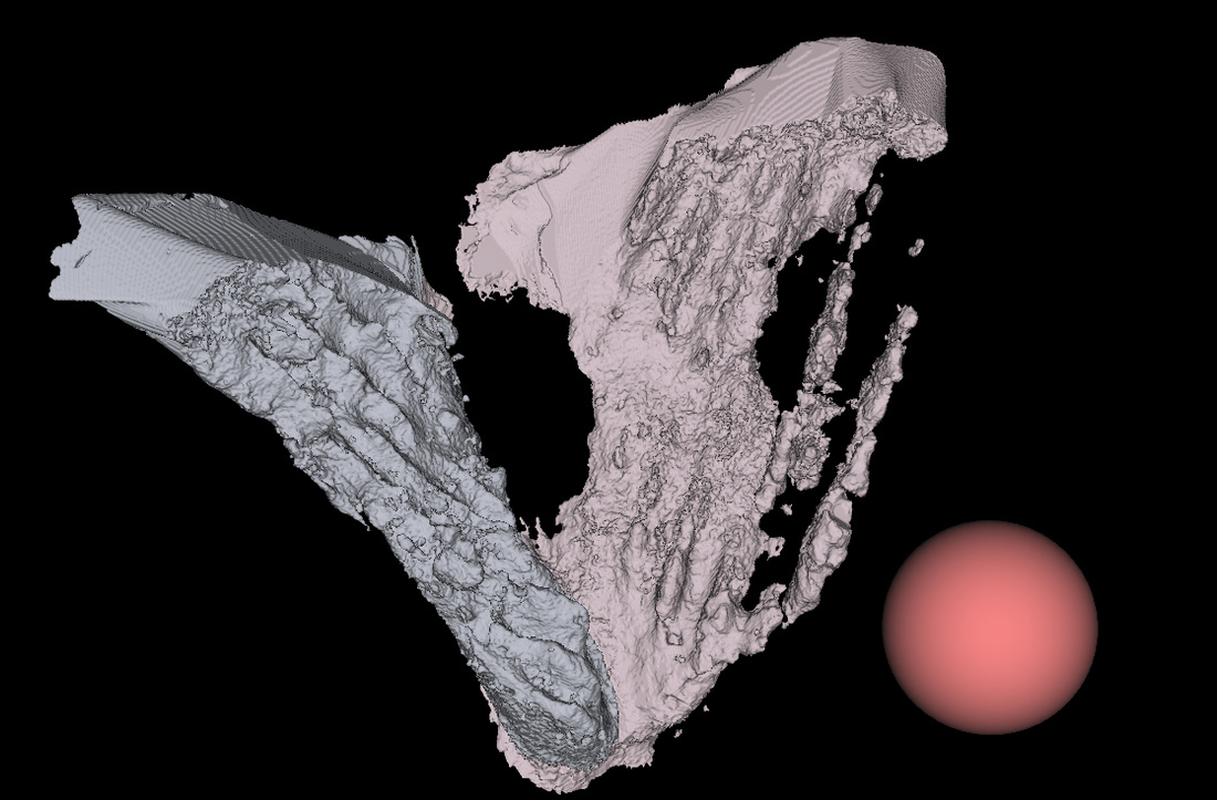
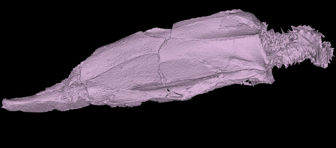
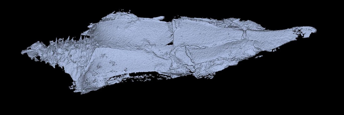
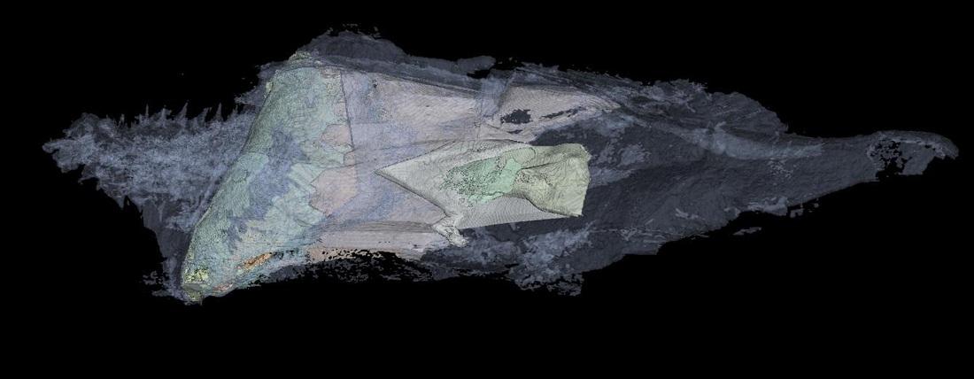
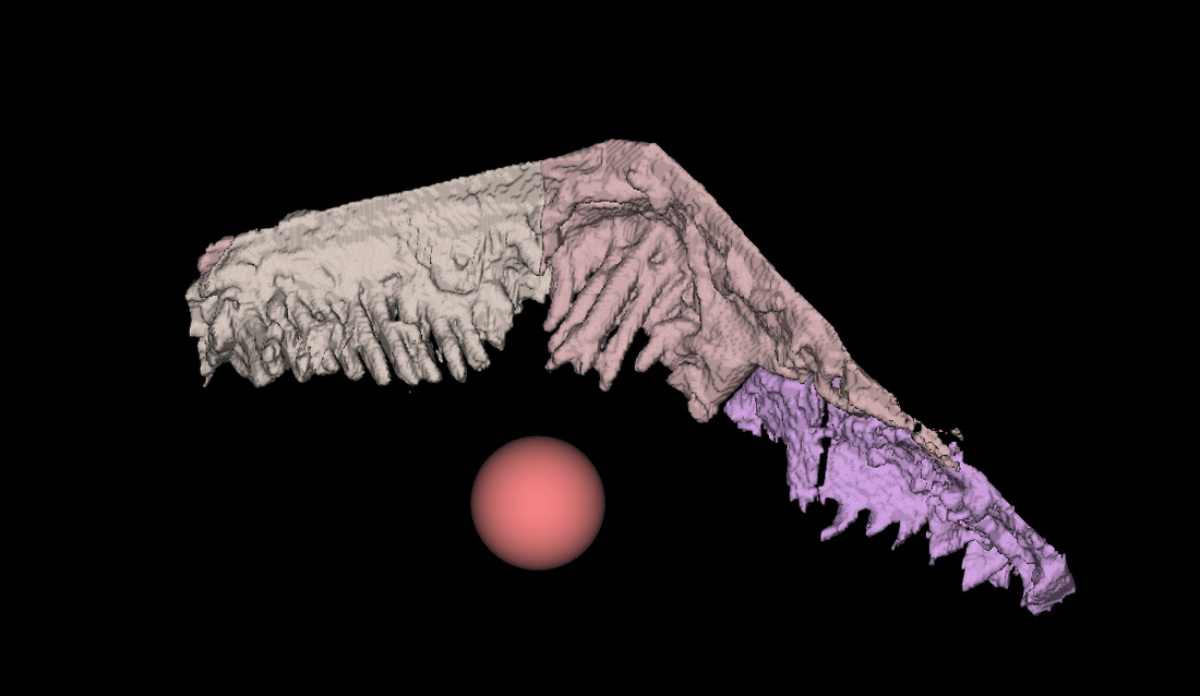
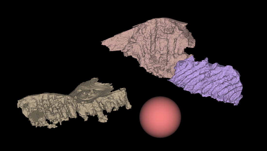
All specimens of Lagynocystis pyramidalis (Stylophora: Mitrata), Darriwilian (Middle Ordovician) in age, belong to the echinoderm collection of the Natural History Museum, London (NHMUK E29453, E29043 and E16107). These fossils were originally collected from the Šárka Formation, Czech Republic (Prague Basin, Central Bohemia). The specimens are preserved as three-dimensional uncompressed, articulated moulds in siliceous nodules, which had been previously split for analysis. Nodules were mounted in florist foam and secured with cello tape to avoid movement during the scanning process.
Three specimens of extant deuterostomes; Balanoglossus sp (Hemichordata: Enteropneusta), Schizocardium sp. (Hemichordata: Enteropneusta), and Branchiostoma floridae (Cephalochordata: Leptocardii), were obtained from the collections of the Université de Montréal (Canada). Specimens were preserved in 70% ethanol (C2H6O). Samples were stained with phosphotungstic acid (PTA) over a period of 17 days in order to enhance the contrast between the collagen of the gill bars and surrounding tissues (Metscher 2009). Specimens were carefully dried, wrapped in cling film or thick foam paper (depending on size) and mounted in plastic graduated cylinders. A piece of PTA soaked foam paper was located at the base of each graduated cylinder to prevent desiccation of the specimens. Tops of graduated cylinders were carefully wrapped with cling film and cello tape to avoid movement during the scanning process.
Hemichordate, cephalochordate and stylophoran specimens were scanned using the X-ray micro-tomography facilities (start-of-the-art Metris X-Tek HMX ST 225 CT scan) at the Natural History Museum, London (NHMUK). Schizocardium sp. was scanned without a filter, voltage kV=150 and 3142 projections. L. pyramidalis (exterior; NHMUK E29453 a_b) was scanned with a copper filter, 0.500mm in thickness, voltage kV=180 and 3142 projections. L. pyramidalis (interior; NHMUK E29453 c) was scanned with a copper filter, 0.250mm in thickness, voltage kV=180 and 3142 projections. B. floridae was scanned without a filter, voltage kV=150 and 3142 projections. Balanoglossus sp. was scanned without a filter, voltage kV=150 and 3142 projections. L. pyramidalis (NHMUK E29043) was scanned with a copper filter, 0.500 mm in thickness, voltage kV=180 and 3142 projections. L. pyramidalis (NHMUK E16107) was scanned with a copper filter, 0.500 mm in thickness, voltage kV=180 and 3142 projections. Stacked images were saved in TIFF format.
Cryptoschisma sp. (MGM-3383D) was scanned using X-ray synchrotron tomographic microscopy in the TOMCAT-Xo2DA (TOmographic Microscopy and Coherent rAdiology experimenTs) beamline at the Paul Scherrer Institute, Zürich. Beamline settings were the same for both specimens ; FE-filter at 50%, filter 1 composed of Aluminium and thickness of 100µm, filter 2 composed copper and thickness of 40µm and filter 3 composed of iron and thickness of 10µm. Beam voltage was keV=36.994. The number of projections for Cryptoschisma sp. is 1201. Stacked images were saved in BMP format.
